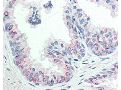
Biomol - antibodies, biochemicals
you have the vision - we have the substance.
Biomol antibodies
deutsche Version english version
Tel: 0800-2466651
 

Category:    Modification: Help finding antibodies

Name/Antigen CLONE HOST TYPE ISOTYPE SPEC. REACT. FORMAT APPLICATION PRODUCER CAT # SIZE PRICE


Antibody results 1
Anti-phospho-AKT (Ser473), clone 17B6.B11.A12.C3.G7.G12 17B6.B11 A12.C3 G7.G Mouse
> sec. Ab
Mab IgG1
kappa
human, mouse

expected: rat, chimpanzee
View abbreviation list.
purified ELISA
WB
IHCView abbreviation list.
Rockland
Icon Datasheet
200-301-268 100 µg 299 €
add to shopping cart
Immunogen: synthetic peptide corresponding to residues surrounding Ser473 of human AKT1 protein

MW: 56 kD

AKT is a component of the PI-3 kinase pathway and is activated by phosphorylation at Ser 473 and Thr 308. AKT is a cytoplasmic protein also known as AKT1, Protein Kinase B (PKB) and rac (related to A and C kinases).

view datasheet
Cat #:
200-301-268
Size:
100 µg
Price:
299 €

UniProt ID: P31749 (Protein knowledgebase)

Antibody Conjugation Kits


Concentration: 1.0 mg/ml (by UV absorbance at 280 nm)
Physical State: Liquid (sterile filtered)
Product Category: Primary Antibodies, Phospho Specific Antibodies
 
Label: Unconjugated
 
Host: Mouse
Clone: 17B6.B11
Immunogen: This monoclonal antibody was produced by repeated immunizations with a synthetic peptide corresponding to residues surrounding S473 of human AKT1 protein.
Gene: AKT1
Reactivity: human, mouse, rat, monkey
Buffer: 0.02 M Potassium Phosphate, 0.15 M Sodium Chloride, pH 7.2
Stabilizer: None
Storage Conditions: Store vial at -20° C prior to opening.  This product is stable for several weeks at 4° C as an undiluted liquid.  Dilute only prior to immediate use.  For extended storage aliquot contents and freeze at -20° C or below.  Avoid cycles of freezing and thawing.  Expiration date is one (1) year from date of opening product.
Synonyms: RAC-PK-alpha, Protein kinase B, PKB, C-AKT
Preservative: 0.01% (w/v) Sodium Azide
 
Background: AKT is a component of the PI-3 kinase pathway and is activated by phosphorylation at Ser 473 and Thr 308. AKT is a cytoplasmic protein also known as AKT1, Protein Kinase B (PKB) and rac (related to A and C kinases). AKT is a key regulator of many signal transduction pathways. AKT Exhibits tight control over cell proliferation and cell viability. Overexpression or inappropriate activation of AKT is noted in many types of cancer. AKT mediates many of the downstream events of PI 3-kinase (a lipid kinase activated by growth factors, cytokines and insulin). PI 3-kinase recruits AKT to the membrane, where it is activated by PDK1 phosphorylation. Once phosphorylated, AKT dissociates from the membrane and phosphorylates targets in the cytoplasm and the cell nucleus. AKT has two main roles: (i) inhibition of apoptosis; (ii) promotion of proliferation.
Application Notes: This monoclonal antibody is suitable for ELISA, immunohistochemistry, immunoprecipitation and western blotting. Expect a band approximately 56 kDa in size corresponding to phosphorylated AKT protein by western blotting in the appropriate cell lysate or extract. This phospho-specific monoclonal antibody reacts with human and mouse AKT pS473 and shows minimal reactivity by ELISA against the non-phosphorylated form of the immunizing peptide. Specific conditions for reactivity should be optimized by the end user. For immunohistochemistry use formalin-fixed paraffin-embedded sections. No pre-treatment of sample is required.
Methods: ELISA
Western Blot
Immunohistochemistry
Immunofluorescence Microscopy
Purity and Specificity: This product was purified from concentrated tissue culture supernate by Protein A chromatography. This antibody is specific for human and mouse AKT protein phosphorylated at S473. A BLAST analysis was used to suggest cross-reactivity with AKT pS473 from human, mouse, rat and chimpanzee sources based on 100% homology with the immunizing sequence. Cross-reactivity with AKT from other sources has not been determined. Cross-reactivity with AKT2 and AKT3 has not been determined.
Recommended Dilutions:
ELISA: 1:20,000
WB: 1:500 - 1:3,000
IHC: 20 µg/ml
IF: 1:500 - 1:3,000
OTHER: User Optimized
General References: Lawlor, M. A. and Alessi, D.R. (2001). PKB/AKT: a key mediator of cell proliferation, survival and insulin responses. J. Cell Science 114:2903-2910.

Alessi, D. R. (2001). Discovery of PDK1, one of the missing links in insulin signal transduction. Biochem. Soc. Trans. 29,1 -14.

Jones,P.F., Jakubowicz,T., Pitossi,F.J., Maurer,F. and Hemmings,B.A. (1991) Molecular cloning and identification of a serine/threonine protein kinase of the second-messenger subfamily. Proc. Natl. Acad. Sci. U.S.A. 88 (10), 4171-4175.


back

More antibodies reacting with this protein:


Biomol Newsletter

Biomol releases hundreds of new products each month.
Stay updated - Register for Biomol's monthly newsletter and receive a 10 EUR voucher for your next purchase at Biomol.
Register now


Prices for antibodies are net excl. shipping and coolant costs as well as VAT.
1    


datasheet datasheet
manual manual
new new
related products related products
 order order


Info: Monthly Special: 50% Discount
Monthly Special: 50% Discount
Info: New Epitomics Rabbit Monoclonal Antibodies since April 2010
New Epitomics Rabbit Monoclonal Antibodies since April 2010
Info: DyLight 488 Conjugated Rabbit Monoclonal Antibodies
DyLight 488 Conjugated Rabbit Monoclonal Antibodies
Info: Bethyl: DyLight(R) and Cy-Conjugated Secondary Antibodies
Bethyl: DyLight(R) and Cy-Conjugated Secondary Antibodies
Info: New Bethyl Products
New Bethyl Products
Info: Antibodies to Proteins in the Anaphase-Promoting Complex/Cyclosome
Antibodies to Proteins in the Anaphase-Promoting Complex/Cyclosome
Info: Save 10% with Cayman
Save 10% with Cayman
Info: New Imgenex Antibodies
New Imgenex Antibodies
Info: Antibodies for the Study of DNA Methylation
Antibodies for the Study of DNA Methylation

show all hits (273 INFOS)


Biomol GmbH
Waidmannstr. 35
22769 HAMBURG
info@biomol.de
www.biomol.de
Tel:+49-40 - 85 32 600
0800 - 24 66 651 free call
Fax: +49-40 - 85 32 60-22
0800 - 24 66 652 free call